|
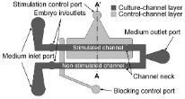
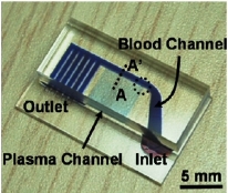
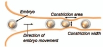
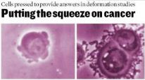
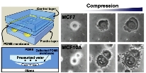
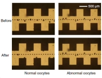
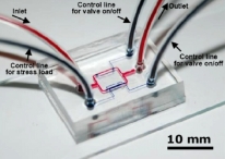
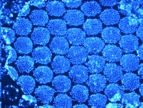
Biomechanical Microdevices for Cell Research The physical forces to which living cells are most commonly exposed are fluid shear, pressure, and stretch. These mechanical stimulations influence the physiological and pathological condition of the organism, which induces many aspects of human health and disease. Cancer cells are also known to have a less extensive internal cytoskeleton than healthy cells, so behave differently when squeezed. We have developed a new kind of microfluidic biomechanical device for compressive stimulation and lysis of cells [Sens. Actuators B Chem. 2007, 128, 108]. Here mechanical stress was applied to the cells with the deflection of the poly(dimethylsiloxane) (PDMS) membrane between two microchannels, formed by multilayer soft lithography. This technology can also be used to spot the difference between cancerous cells and healthy ones by squeezing them until they deform - a discovery that could lead to a cheap tool for cancer detection [Analyst 2008, 133, 1432]. Recently, we also demonstrate a novel microfluidic in vitro cultivation system for bovine embryos that improves their development using a partially constricted channel that mimics peristaltic muscle contraction [Electrophoresis 2009, 30, 3276]. Related Articles: |
||
|
8 |
|
Chae Yun Bae, Minseok S. Kim, Je-Kyun Park, "Mechanical stimulation of bovine embryos in a microfluidic culture platform," Biochip J., 5 (2), 106-113 (2011). |
|
7 |
|
Yu
Chang Kim, Seung-Hoon Kim, Duckjong Kim, Sang-Jin Park, Je-Kyun Park, "Plasma
extraction in a capillary-driven microfluidic
device using surfactant-added poly(dimethylsiloxane)," Sens. Actuators B
Chem.,
145 (2), 861-868 (2010).
|
|
6 |
|
Minseok S. Kim, Chae
Yun Bae, Gabbine Wee, Yong-Mahn Han, Je-Kyun Park, "A
microfluidic in vitro cultivation system for mechanical stimulation of bovine embryos,"
Electrophoresis, 30 (18), 3276-3282
(2009).
|
|
5 |
|
|
|
4 |
|
Yu
Chang Kim, Sang-Jin Park, Je-Kyun Park,
"Biomechanical
analysis of cancerous and normal cells based on bulge
generation in a microfluidic device,"
Analyst, 133 (10), 1432-1439 (2008).
|
|
3 |
|
Wonjae
Choi, Ji-Su Kim, Do-Hyun Lee, Kyung-Kwang Lee, Deog-Bon Koo,
Je-Kyun Park, "Dielectrophoretic oocyte selection chip for in vitro
fertilization,"
Biomed.
Microdevices, 10 (3), 337-345
(2008).
|
|
2 |
|
Yu Chang Kim, Joo H. Kang, Sang-Jin Park, Eui-Soo Yoon, Je-Kyun Park, "Microfluidic biomechanical device for compressive cell stimulation and lysis," Sens. Actuators B Chem., 128 (1), 108-116 (2007). |
|
1 |
|
Minseok
S. Kim, Janghwan Kim, Hyo-Won Han, Yee Sook Cho, Yong-Mahn
Han, Je-Kyun Park,
"Microfabricated
embryonic stem cell divider for large-scale propagation of
human embryonic stem cells," Lab Chip,
7 (4), 513-515 (2007).
|
|
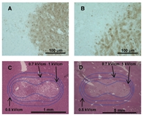
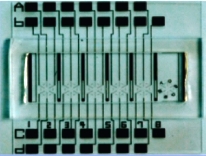
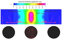
Microfabricated Devices for Electrochemotherapy A microfabricated cell-based electrochemotherapy (ECT) testing device which mimics a clinical electroporator of circular needle-array is demonstrated to study the electrochemotherapeutic effect on T47D human breast cancer cells. Until now, the performance between electroporators having two- and six-needle circular array electrodes, which are the general needle-type clinical electroporators for ECT, has not been evaluated systemically, although many studies have investigated the efficacy of ECT on cancer cells. In this study, the cell-based performance on the newly developed ECT testing device was analyzed in two and six-electrode modes using propidium iodide and bleomycin, and the electroporation characteristics were characterized [Biomed. Microdevices 2009, 11, 151; Anal. Chem. 2009, 81, 3517]. We also developed a microfabricated electroporator for the irreversible electroporation (IRE) of tissues by miniaturizing a clinical electroporator with a two-needle array while keeping the same electric field strength distribution. With the developed microfabricated electroporator, the effect of IRE on rat liver tissues was analyzed with time by immunohistological stainings and electrical measurement, and the experimental results were compared with those operated with the corresponding real-scale clinical electroporator [Tissue Eng. Part C Methods 2010, 16, 1245]. Related Articles: |
||
|
3 |
|
Youn-Suk Choi, Hong-Bae
Kim, Junho Chung, Hyung-Sik Kim, Jeong-Han Yi, Je-Kyun Park, "Preclinical
analysis of irreversible electroporation on rat liver tissues using a microfabricated
electroporator,"
Tissue Eng. Part C Methods, 16
(6), 1245-1253 (2010). |
|
2 |
|
Youn-Suk Choi, Hong-Bae
Kim, Seung-Hoon Kim, Jaekyu Choi, Je-Kyun Park, "Microdevice
for analyzing the effect of electrochemotherapy on cancer cells," Anal.
Chem.,
81 (9),
3517-3522 (2009). |
|
1 |
|
Youn-Suk Choi, Hong-Bae Kim, Gil-Sik Kwon, Je-Kyun Park, "On-chip testing device for electrochemotherapeutic effects on
human breast cells," Biomed.
Microdevices, 11 (1), 151-159 (2009).
|